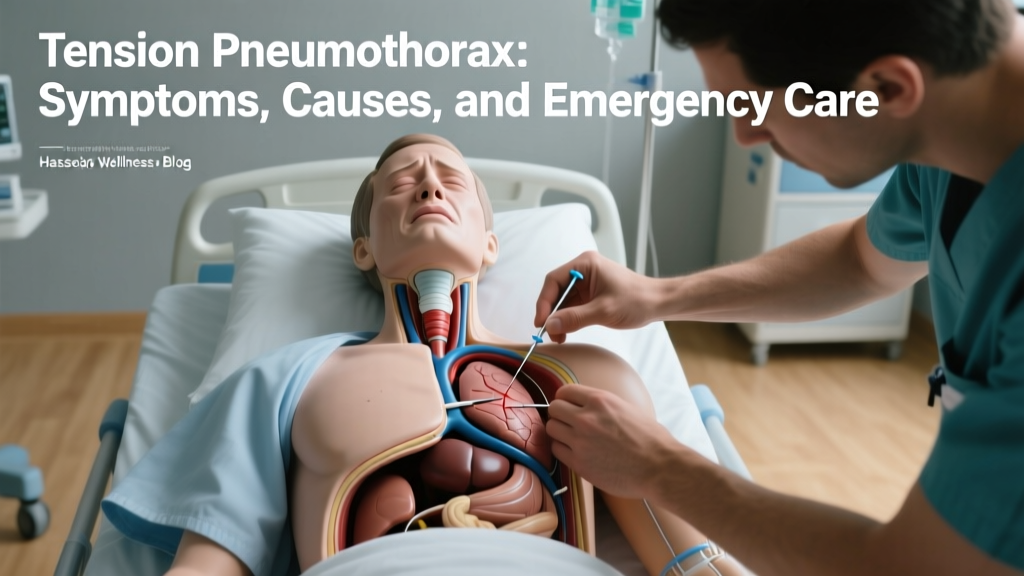
A tension pneumothorax is a life-threatening medical emergency that occurs when air enters the pleural space—the cavity between the lung and the chest wall—but cannot escape. This trapped air creates a one-way valve effect, causing intrapleural pressure to rise rapidly. As pressure builds, it compresses the affected lung and pushes the mediastinum (the central compartment of the chest containing the heart and major blood vessels) toward the opposite side. This mediastinal shift severely restricts venous return to the heart, leading to a catastrophic drop in cardiac output and obstructive shock. Understanding the mechanics and immediate interventions for this condition is critical for first responders, medical students, and anyone seeking to comprehend acute thoracic trauma.
The Pathophysiology: How a Tension Pneumothorax Develops
To understand the severity of this condition, you must first grasp the normal mechanics of breathing. The lungs are surrounded by a double-layered membrane called the pleura. A thin layer of lubricating fluid between these layers creates negative pressure, keeping the lungs expanded against the chest wall.
When a puncture occurs—whether from a fractured rib, a penetrating wound, or a ruptured bleb (a small air sac on the lung)—air breaches this space. In a simple pneumothorax, air enters and equalizes with atmospheric pressure. In a tension pneumothorax, the damaged tissue flap acts as a one-way valve. Air is forced into the pleural cavity during inhalation but is trapped during exhalation. With every breath, intrapleural pressure exceeds atmospheric pressure, collapsing the ipsilateral lung and compressing the contralateral lung and heart. According to the National Center for Biotechnology Information (NCBI), this progressive pressure buildup is what transitions the condition from a localized respiratory issue to a systemic cardiovascular collapse.
Clinical Presentation: Identifying the Warning Signs
Time is tissue. Recognizing the clinical signs before cardiovascular collapse is paramount. Medical professionals rely on a specific cluster of physical findings rather than waiting for radiographic confirmation.
Cardinal Signs of Obstructive Shock
- Severe Respiratory Distress: Rapid, shallow breathing (tachypnea) and a desperate sensation of air hunger.
- Tracheal Deviation: The trachea shifts away from the affected side due to the massive pressure buildup pushing mediastinal structures to the opposite hemithorax. This is a late and highly critical sign.
- Distended Neck Veins: Jugular venous distension (JVD) occurs because the shifted mediastinum compresses the superior vena cava, preventing blood from draining from the head and neck back to the right atrium.
- Unilateral Absent Breath Sounds: The collapsed lung produces no audible air movement on the affected side when auscultated with a stethoscope.
- Hypotension and Tachycardia: The heart beats faster to compensate for the dropping blood pressure caused by decreased venous return.
- Hyperresonance to Percussion: Tapping on the chest wall yields a drum-like sound, indicating trapped air rather than healthy lung tissue or fluid.
Simple vs. Tension Pneumothorax: A Clinical Comparison
Distinguishing between a simple collapsed lung and a tension emergency dictates the speed and aggression of the medical response. The Cleveland Clinic notes that while both involve air in the pleural space, the hemodynamic consequences are vastly different.
| Clinical Feature | Simple Pneumothorax | Tension Pneumothorax |
|---|---|---|
| Intrapleural Pressure | Equal to or slightly above atmospheric | Progressively higher than atmospheric |
| Mediastinal Shift | None or minimal | Severe shift to the contralateral side |
| Hemodynamic Status | Stable blood pressure and heart rate | Hypotension, tachycardia, obstructive shock |
| Tracheal Position | Midline | Deviated away from the affected side |
| Primary Intervention | Observation, supplemental oxygen, or chest tube | Immediate needle decompression followed by chest tube |
Primary Etiologies and Risk Factors
Traumatic Origins
Blunt or penetrating chest trauma is the most common cause. Motor vehicle collisions, stab wounds, and gunshot wounds can tear the visceral or parietal pleura. Iatrogenic (medically induced) trauma also accounts for a significant percentage of cases, often resulting from central venous catheter placement, thoracentesis, or barotrauma from mechanical ventilation in ICU settings.
Spontaneous Ruptures
While less likely to progress to a tension state than traumatic injuries, spontaneous pneumothoraces can become tension emergencies if a one-way valve forms. This typically happens when subpleural blebs or bullae rupture, often in tall, thin young males or patients with underlying lung diseases like COPD, asthma, or cystic fibrosis.
Diagnostic Modalities: Beyond the Stethoscope
While clinical diagnosis dictates immediate treatment, adjunct diagnostic tools are vital once the patient is stabilized or if the presentation is ambiguous.
Extended Focused Assessment with Sonography for Trauma (eFAST)
Point-of-care ultrasound (POCUS) has revolutionized thoracic trauma assessment. In a healthy lung, the sliding of the visceral and parietal pleura against each other creates a "lung sliding" artifact on ultrasound. The absence of lung sliding, combined with the presence of a "barcode sign" or "stratosphere sign" on M-mode ultrasound, is highly sensitive for pneumothorax. Ultrasound is rapid, non-invasive, and does not expose the patient to ionizing radiation.
Radiography and Computed Tomography
A standard upright chest X-ray will reveal a lack of lung markings and a visible visceral pleural edge. However, in a true tension emergency, obtaining an X-ray delays life-saving decompression. Computed Tomography (CT) of the chest is the gold standard for diagnosing small or loculated pneumothoraces and identifying underlying structural lung disease, but it is strictly reserved for hemodynamically stable patients.
Emergency Interventions: Decompression Protocols
A tension pneumothorax is a clinical diagnosis, not a radiological one. Waiting for a chest X-ray can be fatal. Immediate decompression is required to convert the tension pneumothorax back into a simple pneumothorax, restoring venous return and cardiac output.
Needle Thoracostomy (Needle Decompression)
This is the immediate bridging intervention. A large-bore (14-gauge or 16-gauge) needle is inserted into the pleural space to vent the trapped air. According to the Advanced Trauma Life Support (ATLS) guidelines, the traditional insertion site is the second intercostal space in the midclavicular line. However, recent evidence and updated ATLS protocols frequently recommend the fourth or fifth intercostal space in the anterior axillary line. This lateral approach is often preferred because chest wall thickness is typically lower here, reducing the risk of the needle failing to reach the pleural cavity in patients with higher body mass.
Tube Thoracostomy (Chest Tube Insertion)
Needle decompression is strictly temporary. Definitive management requires the insertion of a chest tube connected to an underwater seal drainage system. The tube is typically placed in the "safe triangle" (bordered by the anterior border of the latissimus dorsi, the lateral border of the pectoralis major, a line superior to the horizontal level of the nipple, and an apex below the axilla). This allows continuous evacuation of air and gradual re-expansion of the lung. As noted by MedlinePlus, the chest tube remains until the air leak seals and the lung fully adheres to the chest wall again.
Prognosis and Post-Acute Management
Once the chest tube is placed and the lung re-expands, the immediate threat to life resolves. The chest tube usually remains in place for 2 to 5 days until air leakage ceases entirely. Patients with spontaneous origins may require surgical interventions like chemical pleurodesis or mechanical bullectomy to prevent recurrence. The risk of a second spontaneous pneumothorax is roughly 30% to 50% after the first episode, making surgical consultation a standard part of post-acute care for non-traumatic cases.